Tumor visualization
Imaging plays a crucial role in visualizing the tumor, surrounding organs, and
the patient’s anatomy. By obtaining high-quality and detailed images, we can
precisely identify the size, shape, and location of the tumor within the
patient’s body, enabling us to accurately target it. The information derived
from these images guides us in determining the optimal radiation dosage and the
most effective delivery method.
Treatment planning
Every treatment plan is tailored to the patient’s unique requirements, outlining
the precise amount of radiation that the cancer cells should receive and the
angles from which it will be administered. A treatment schedule will also be
developed during this phase. The added advantage Ethos offers is the real-time,
on-couch adaptation that enables last minute customization of the treatment
plan.
Treatment delivery
During the treatment session, the radiation therapist will assist you in
positioning the patient comfortably on the treatment table. Just before the
treatment begins, images will be taken to enable the care team to verify the
precise size and location of the tumor. After the necessary adjustments to the
treatment plan are made based on these images, treatment will commence.
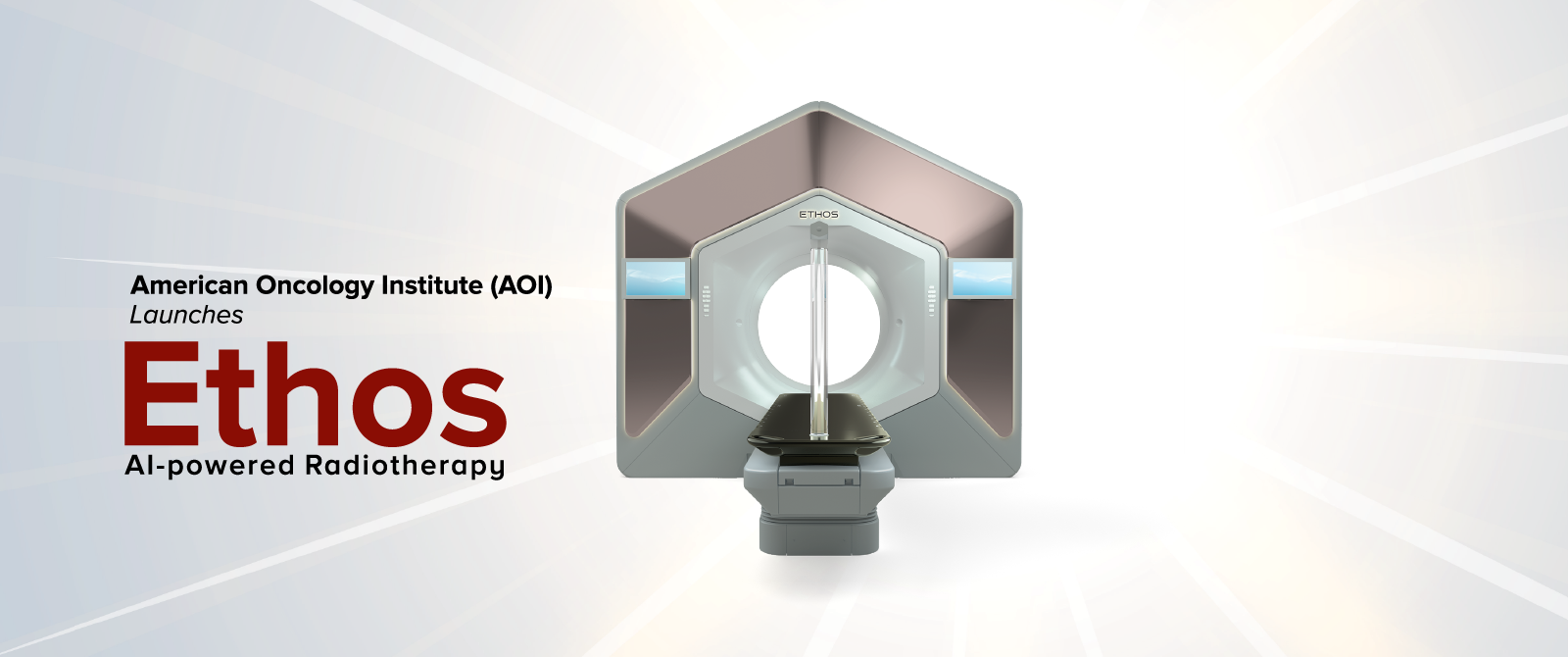
The radiation is delivered using a linear accelerator (linac), a machine that
revolves around the patient while projecting the beam. With Ethos™ therapy, the
rotating beam gantry is enclosed within the opening (the bore), so the patient
won't perceive any visible rotation.
Throughout the treatment, the patient will maintain continuous communication with
their therapist via an integrated camera system and a two-way intercom. While
the first treatment session may take a little longer due to additional setup
requirements, the entire process typically lasts just a few minutes.